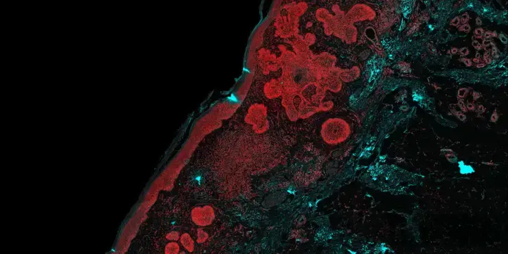
illumiSonics Announces Publication in Diagnostics Showing Strong Diagnostic Concordance Between PARS® Label-Free Virtual H&E and Conventional H&E Staining in Skin Cancer
Seven fellowship-trained dermatopathologists found virtual H&E images generated from unstained tissue to be highly concordant with gold-standard chemical staining
Waterloo, ON – October 14, 2026 – illumiSonics Inc. today announced the publication of a new peer-reviewed study in Diagnostics evaluating the diagnostic performance of virtual hematoxylin and eosin (H&E) images generated with Photon Absorption Remote Sensing (PARS®) in skin cancer excisional biopsies. The study found that PARS virtual H&E images performed equivalently to conventional chemically stained H&E slides across diagnosis, subtyping and surgical margin assessment.
The paper, titled “Photon Absorption Remote Sensing Virtual Histopathology: A Preliminary Exploration of Diagnostic Equivalence to Gold-Standard H&E Staining in Skin Cancer Excisional Biopsies,” is open access and was led by researchers at the University of Waterloo in collaboration with clinicians and scientists at the University of Calgary, the University of Toronto and Indiana University School of Medicine.
In the study, sixteen unstained formalin-fixed paraffin-embedded skin excisions, including squamous cell carcinoma (SCC), basal cell carcinoma (BCC) and normal skin, were imaged with PARS and then stained with conventional H&E. Seven fellowship-trained dermatopathologists independently assessed all 32 images (16 PARS and 16 H&E) in a masked, randomized fashion.
Key findings:
Diagnostic performance was comparable between modalities. PARS virtual H&E achieved a sensitivity of 96.7% and specificity of 95.2%, compared with 95.6% and 85.7%, respectively, for chemically stained H&E.
Primary diagnostic agreement between modalities was 95.5%, with high inter-rater reliability among the seven dermatopathologists (Fleiss’ κ = 0.886 for PARS and 0.805 for H&E).
Subtyping agreement was strong. Agreement between modalities was 91.8% for SCC and 100% for BCC.
Margin assessment agreement was 92.2% for margin confinement.
Readers could not reliably distinguish PARS from H&E images, and diagnostic confidence was statistically equivalent between modalities.
PARS is a label-free technology that images tissue by observing optical absorption, and, when coupled with deep learning, produces virtual H&E images without chemical staining. Because the tissue is not stained or destroyed, it remains available for downstream molecular and genomic analysis, and the digital images are well suited to integration with AI-based pathology tools.
“These results are an encouraging step for our technology,” said John Mackey, CEO of illumiSonics Inc. “Dermatopathologists found PARS virtual H&E images to be highly concordant with conventional staining, while the tissue is preserved for further testing. We have launched prospective validation studies to advance our MLI™ platform into the clinic.”
"Achieving over 90% diagnostic agreement with conventional H&E is a huge milestone to translate years of research into the clinic. This study shows that pathologists interpreted our label-free virtual stains with the same confidence as chemical H&E, laying an exciting foundation for large-scale clinical validation,” said Benjamin Ecclestone, first study author and Principal Research Scientist at illumiSonics Inc.
Example PARS and chemical H&E whole-slide images from the study are publicly available through the BioImage Archive (accession S-BIAD2324).This work was supported, in part, by an award from the INOVAIT Focus Fund. illumiSonics is grateful to INOVAIT for its continued support of Canadian medical innovation.
Read the full paper (PDF): https://www.mdpi.com/2075-4418/16/20/3315/pdf
Citation: Ecclestone, B.R.; Tweel, J.E.D.; Abi Daoud, M.; Gaouda, H.; Dinakaran, D.; Wallace, M.P.; Somani, A.K.; Bigras, G.; Mackey, J.R.; Haji Reza, P. Photon Absorption Remote Sensing Virtual Histopathology: A Preliminary Exploration of Diagnostic Equivalence to Gold-Standard H&E Staining in Skin Cancer Excisional Biopsies. Diagnostics 2026, 16(20), 3315.
About illumiSonics Inc.
illumiSonics Inc. is a Waterloo, Ontario based company that has developed the MLI™ (Multi-Laser Imaging) platform: a transformative imaging device for life science research, digital pathology, fresh tissue diagnostics and surgical margin assessment. MLI™ is the most advanced optical absorption imaging device to date. It captures optical absorption from radiative contrast (e.g., autofluorescence), non-radiative relaxation (e.g., temperature, pressure) and scattering channels (indirect optical absorption measurement) simultaneously without contact with the tissue. The ability to non-destructively generate multiple diagnoses from a single fixed or fresh tissue sample without staining addresses an important unmet medical need. For more information, visit www.illumisonics.com.
Media Contact
illumiSonics Inc.
Waterloo, ON, Canada
Phone: +1 226-407-7873
Email: contact@illumisonics.com