Comparison of MLI® and Chemical H&E Staining in Skin Cancer
- Mar 11
- 1 min read
Updated: Apr 8
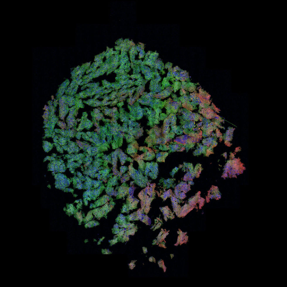
Unenhanced whole slide MLI® of human skin cancer (left panel) demonstrating basal cell carcinoma (BCC) architecture. Tissue and interpretation from Dr. Marie Abi Daoud, University of Calgary.


The first zoom-in shows necrosis within a tumour extending from the epidermis. The second shows an isolated tumour nest in the dermis, along with nearby skin glands. The third shows a higher-magnification view highlighting intranuclear detail within tumor cells, including visible nucleoli. These features demonstrate the ability of MLI® contrast to resolve both large-scale tumor architecture, and fine cellular and stromal features within skin cancer; note the exquisite collagen details. After MLI®, the unstained tissue section was then stained with chemical H&E for direct image comparisons (right panels).


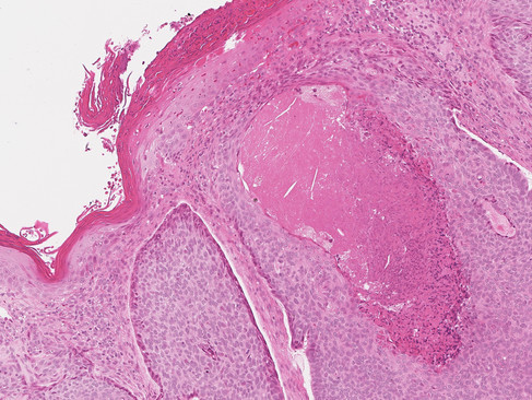


Image Credit: illumiSonics Inc.