MLI™ of Endometriosis
- Mar 12
- 1 min read
Updated: 5 days ago
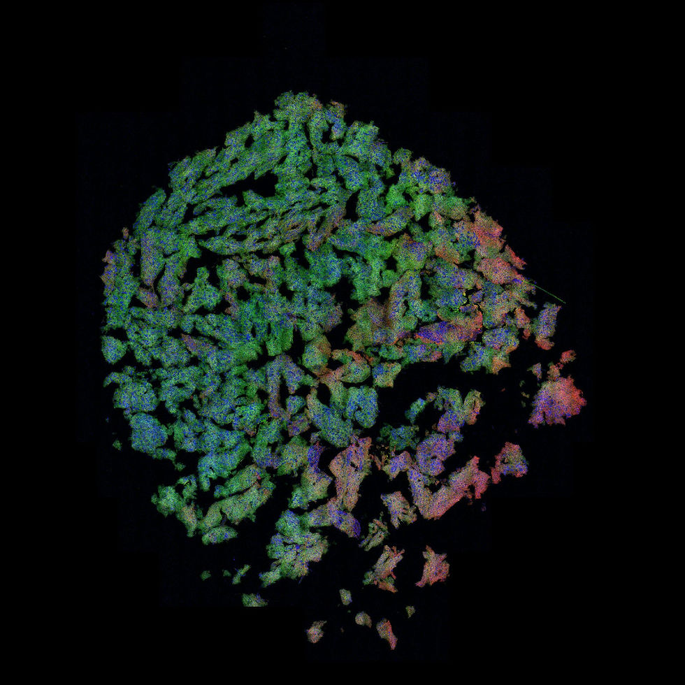
Unenhanced MLI™ of an unstained pelvic biopsy specimen readily identifies areas of endometriosis.


Both the unenhanced MLI™ and H&E-stained photomicrograph demonstrates histologic features consistent with endometriosis in a endoscopic biopsy of pelvic tissue. The section shows ectopic endometrial-type glands with a rim of endometrial stroma embedded in a background of dense fibroconnective tissue. The glands are irregular and elongated, lined by columnar epithelial cells with basally oriented nuclei, resembling endometrial glands.
Surrounding the glands is endometrial-type stroma composed of small, uniform spindle cells, which supports the diagnosis of endometriosis when present together with glandular elements. The adjacent tissue demonstrates fibrosis.
Image Credit: illumiSonics Inc.