MLI® of Breast Ductal Carcinoma in Situ (DCIS)
- Mar 10
- 1 min read
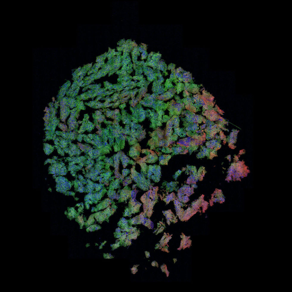
Unenhanced MLI® alongside MLI® virtual H&E showing the diagnostic features of human breast ductal carcinoma in situ. Tissue courtesy of Ontario Institute for Cancer Research.


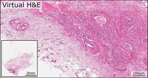
Both the unenhanced MLI® together with the MLI® virtual H&E demonstrate the diagnostic features of ductal carcinoma in situ (DCIS) of the human breast. The section shows markedly distended ductal structures filled with a proliferation of atypical epithelial cells. These neoplastic cells form solid and cribriform architectural patterns, with irregular secondary lumina visible within the intraductal proliferation. The malignant cells exhibit increased nuclear density, hyperchromasia, and loss of normal epithelial polarity. The involved ducts remain bounded by an intact basement membrane and surrounding fibrous stroma, indicating that the lesion is confined within the ductal system without stromal invasion. In several areas there is central luminal debris showing comedo-type necrosis. The surrounding stroma demonstrates reactive fibrous tissue and mild inflammatory infiltrate.
Image Credit: Tissue courtesy of OICR, images by illumiSonics Inc.