Comparison of MLI®, MLI® Virtual H&E, and H&E in Invasive Breast Cancer
- Mar 6
- 1 min read
Updated: Mar 24
MLI® whole slide of invasive ductal carcinoma. Tissue courtesy of Ontario Institute for Cancer Research.


The above three whole slide images show raw MLI® contrast and MLI® virtual H&E derived from unstained tissue, and the same tissue after H&E chemical staining.


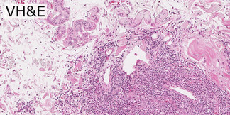


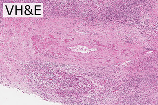


Zoom-ins highlight the complex data captured in the raw MLI® contrast in unstained tissue, and the excellent concordance between MLI® virtual H&E, paired with the same tissue subsequently stained with chemical H&E.
The lower right of the tissue shows invasive breast carcinoma with a prominent tumor-associated lymphocytic response. Irregular malignant glands and small clusters of epithelial tumor cells infiltrate a fibrous desmoplastic stroma, consistent with invasive carcinoma. The upper left of the tissue shows normal ductal elements.
Note the complex stromal detail captured by the raw MLI® in the leftmost panels: much of this information is not evident in H&E stained tissue.
Image Credit: Tissues courtesy of OICR, images by illumiSonics Inc.


