MLI™ of Ocular Tissue
Updated: Jul 16
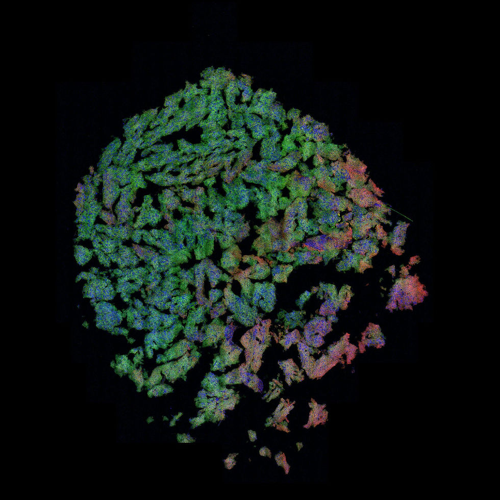
In collaboration with the research team led by Prof. Kathleen Hill [Biology, Western University], we are exploring how Multi-Laser Imaging (MLI™) can reveal rich biological information from unstained tissue. By capturing multiple complementary intrinsic biological signals in a single, non-destructive scan, MLI™ provides detailed views of tissue architecture while preserving samples for future analyses. The images below showcase representative retinal scans acquired from unstained tissue, demonstrating the rich structural and molecular information that can be visualized without dyes or labels.
MLI™ Whole Slide - 1 MLI™ Whole Slide - 2
Intrinsic Contrast in Ocular Anatomy
Relying entirely on innate tissue properties, MLI™ utilizes distinct contrast mechanisms to map complex ocular anatomy simultaneously:
Non-Radiative Contrast: Provides exceptionally strong visualization of the nuclear layers, clearly highlighting cellular density and structural boundaries.
Radiative Autofluorescence: Complements the nuclear detail by capturing the photoreceptor layers and surrounding supporting tissue compartments.
Eye 1 - Zoom 1 Eye 2 - Zoom 1 Eye 1 - Zoom 2 Eye 2 - Zoom 2
Label-Free Structural Assessment
By capturing these intrinsic signals, MLI™ provides a clear structural baseline for observing broad architectural features within the retina. Visualizing the nuclear layers alongside photoreceptor compartments delivers rich microanatomical insights into cellular organization. Because the MLI™ workflow requires no chemical staining, it accomplishes this while leaving the tissue entirely intact for complementary downstream analysis.